Chapter 4 · 19 min
Circuits and an Organized Brain
Chapter 4 of How the Brain Works: Circuits and an Organized Brain.
How the Brain Works | Ch 4 Part 1/2: Circuits and an Organized Brain ↗How the Brain Works | Ch 4 Part 2/2: Circuits and an Organized Brain ↗Visual credits
12 sourcesSources and credits for imagery used in this film, including material incorporated into explanatory animations.
Human brain, sagittal section 1
Themium
View original source ↗Source licence: CC0Incorporated into an explanatory animation; presentation may include cropping, labels and overlays.
Thomas Willis, Cerebri anatome, Figure I (1664)
Thomas Willis; illustration credited in the 1664 preface to Christopher Wren; Wellcome Collection scan
View original source ↗Source licence: CC BY 4.0Incorporated into an explanatory animation; presentation may include cropping, labels and overlays.
Midline sagittal view of the brainstem and cerebellum - Arbor vitae
Christian Brogna, José Pedro Lavrador, Hussein Shaaban Kandeel, Ahmad Beyh, Eduardo C. Ribas, Francesco Vergani & Christos M. Tolias
View original source ↗Source licence: CC BY 4.0Human brain left midsagitttal view closeup description 2
John A Beal, PhD Dep't. of Cellular Biology & Anatomy, Louisiana State University Health Sciences Center Shreveport
View original source ↗Source licence: CC BY 2.5Incorporated into an explanatory animation; presentation may include cropping, labels and overlays.
Human Brodmann cortical fields, 1909
Korbinian Brodmann
View original source ↗Source licence: Public domainIncorporated into an explanatory animation; presentation may include cropping, labels and overlays.
Gray anatomy plate752, exposed association fibres
Henry Vandyke Carter; Henry Gray, revised by Warren H. Lewis
View original source ↗Source licence: Public domainIncorporated into an explanatory animation; presentation may include cropping, labels and overlays.
Hippocampus
Images are generated by Life Science Databases(LSDB).
View original source ↗Source licence: CC BY-SA 2.1 jpIncorporated into an explanatory animation; presentation may include cropping, labels and overlays.
Ran et al. 2022, Figure 1: Tuning of NTS neurons to gut and upper airway stimuli
Chen Ran, Jack C. Boettcher, Judith A. Kaye, Catherine E. Gallori and Stephen D. Liberles
View original source ↗Source licence: CC BY 4.0Incorporated into an explanatory animation; presentation may include cropping, labels and overlays.
Dti-tractography-normal-corpus-callosum
TAbildskov
View original source ↗Source licence: CC BY-SA 3.0Incorporated into an explanatory animation; presentation may include cropping, labels and overlays.
Maier-Hein 2017 Figure 5, complete standalone figure artwork
Klaus H. Maier-Hein and coauthors
View original source ↗Source licence: CC BY4.0Incorporated into an explanatory animation; presentation may include cropping, labels and overlays.
Gordon et al. 2023, Figure 1: Precision functional mapping of primary motor cortex
Evan M. Gordon, Roselyne J. Chauvin, Andrew N. Van and coauthors
View original source ↗Source licence: CC BY 4.0Incorporated into an explanatory animation; presentation may include cropping, labels and overlays.
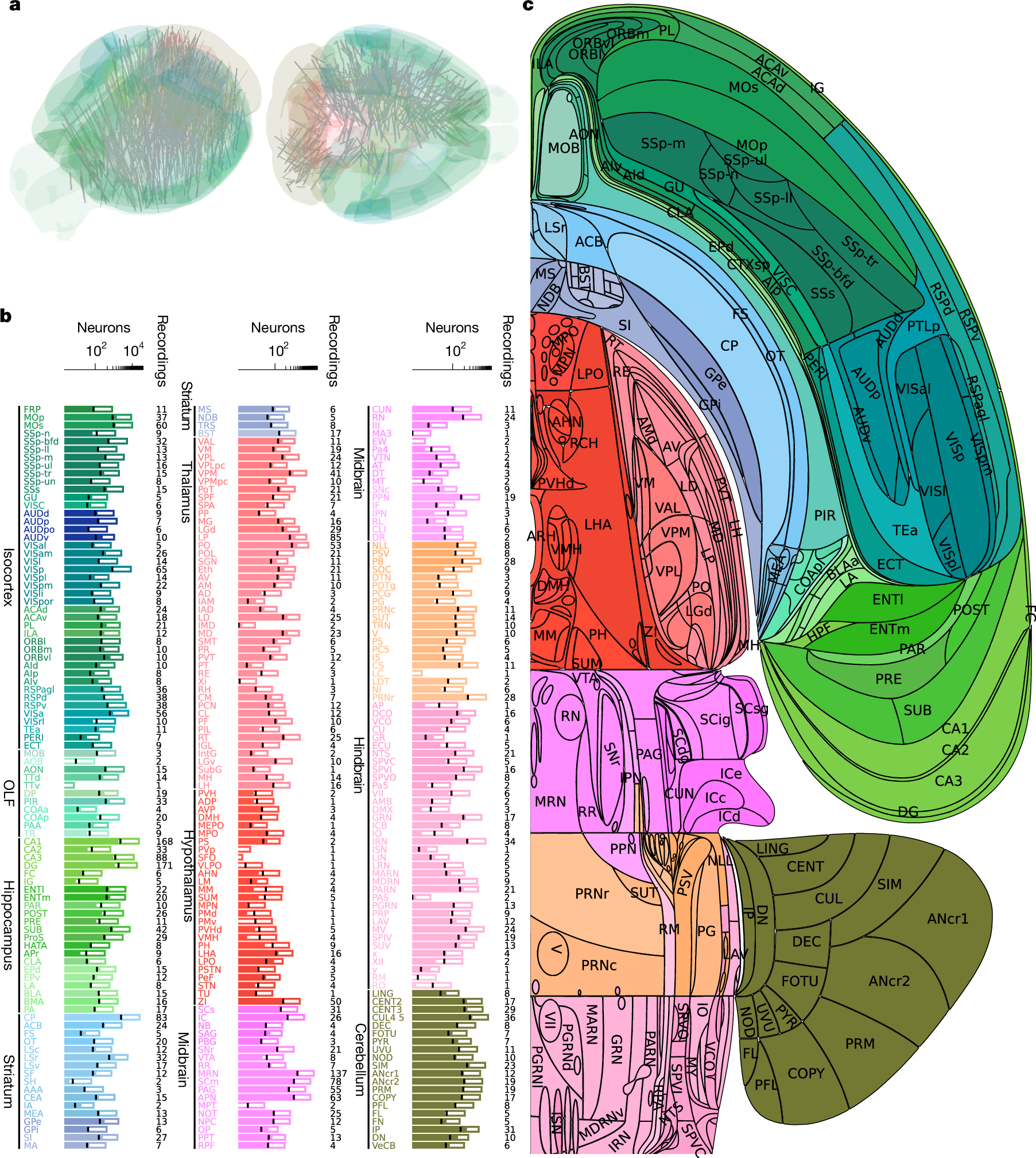
IBL brain-wide recordings, Figure2
International Brain Laboratory and named coauthors
View original source ↗Source licence: CC BY4.0Incorporated into an explanatory animation; presentation may include cropping, labels and overlays.


.jpg)


Research references
Selected sources from the episode’s research and scientific figures.
- Nature paper ↗
- Primary record and DOI ↗
- Dichter et al. human vocal-pitch recording and stimulation ↗
- Schmitt et al. 2017 ↗
- https://www.nature.com/articles/nature11846 ↗
- Azevedo et al. 2009 ↗
- Schmahmann and Sherman 1998 ↗
- Zeng et al., Science, 26 October 2018 ↗
- Kupari et al. 2019 ↗
- Primary paper ↗
- Maier-Hein et al. 2017 ↗
- https://pmc.ncbi.nlm.nih.gov/articles/PMC3174820/ ↗
- https://www.nature.com/articles/s41586-023-05964-2 ↗
- Lavenex and Amaral 2000 ↗
- 2023 primary human study ↗
- Source page and reuse statement ↗
- Version 1 README, p. 1 ↗
- Digitized book, 548 images ↗
- Journey of Sound, NIH/NIDCD ↗
- How Does the Human Body Produce Voice and Speech?, NIH ↗
- Brain-wide map, Sainsbury Wellcome Centre ↗
- Brainwide Map and Beyond, Gaëlle Chapuis / NeuroFM Workshop ↗
- Willis historical context ↗
- https://download.alleninstitute.org/informatics-archive/allen_human_reference_atlas_3d_2020/version_1/README.pdf): ↗
- https://wellcomecollection.org/works/kv8rzxwe): ↗
- https://www.youtube.com/watch?v=JF8rlKuSoFM): ↗
- https://www.youtube.com/watch?v=ETvTSwEgJRM): ↗
- https://royalsociety.org/blog/2022/01/circle-of-willis/): ↗
- NIDCD hearing account ↗
- UT Houston auditory pathways, section 13.1 ↗
- García-Gomar et al. human subcortical MRI atlas ↗
- Simonyan and Horwitz 2011 ↗
- Primary text ↗
- corticofugal gating in anesthetized guinea pigs ↗
- Sherman 2007 ↗
- Hwang et al. 2017 ↗
- Rikhye et al. 2018 ↗
- Mukherjee et al. 2021 ↗
- Scott, Mukherjee, Nassar and Halassa 2024 ↗
- abstract ↗
- https://www.science.org/doi/10.1126/science.adr3675 ↗
- AAAS publisher release ↗
- Purves, Circuits within the Basal Ganglia System ↗
- 2023 beginners review ↗
- Calabresi et al. 2014 ↗
- PMC13323681 ↗
- Schmahmann's 2019 review ↗
- 2021 update ↗
- Herculano-Houzel's 2010 comparative analysis ↗
- Tanaka et al. 2020 ↗
- Hull 2020, eLife 9:e54073 ↗
- 2024 Cerebral Cortex study, bhae382 ↗
- Prescott and Liberles 2022, *Internal senses of the vagus nerve* ↗
- full-text route ↗
- Min et al. 2019 ↗
- 2019 autonomic critique ↗
- Bai et al. 2019, genetic identification of feeding-related vagal sensory neurons ↗
- Chen et al. 2015 ↗
- 2024 arcuate review ↗
- *Hunger from within* 2021 ↗
- Ran et al. 2022, *A brainstem map for visceral sensations* ↗
- Craig 2002 ↗
- 2003 article ↗
- Del Negro, Funk and Feldman 2018 ↗
- PubMed with figures ↗
- *Respiratory rhythm: an emergent network property?* ↗
- Vann et al. 2018 ↗
- Yeo et al. 2011 ↗
- Logothetis et al. 2001, monkey physiology/fMRI comparison ↗
- IBL 2025 ↗
- Author-institution record ↗
- author-hosted primary PDF ↗
- Schilling et al. 2019 ↗
- 2019 benchmark review ↗
- Author release ↗
- Honey et al. 2009 ↗
- NIMH ↗
- S1200 release ↗
- Primary record ↗
- author-lab full text ↗
- published-version repository ↗
- IBL source-owner page ↗
- Lemon 2008, *Descending Pathways in Motor Control* ↗
- Institutional teaching copy ↗
- author PDF ↗
- institutional record ↗
- Muret, Makin and Diedrichsen's open review ↗
- *White matter connections within the central sulcus subserving the somato-cognitive action network*, Brain 2025 ↗
- Primary paper ↗
- author-lab record ↗
- Lavenex, Suzuki and Amaral 2004 ↗
- plate record ↗
- Project ↗
- dataset license discussion ↗
- CONP dataset ↗
- institutional release ↗
- Young Adult project ↗
- IBL policies ↗
- PMC ↗
- What Is Sound?, NIH4Health ↗
- Hearing: A Gateway to Our World, NIH ↗
- Data from IBL, Neurodata Without Borders ↗
- IBL dataset, Neuromatch ↗
- search hit ↗
How this film was made
Created and editorially directed by Anders Lunde using a custom production system developed over six months. AI assists research, drafting, programming, and visual production; I review and edit the material and make the final editorial decisions. Narration uses text-to-speech. Visuals combine custom-built scientific animations, AI-generated reconstructions, and credited scientific imagery.
Generated reconstructions illustrate scientific interpretations. They are distinct from photographs, specimens, and measured data.
More about the creator and production →